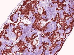
During immunohistochemistry (IHC) staining, proteins are observed under a brightfield microscope by employing enzymatic markers, like DAB chromogen, to highlight the linked proteins. This staining technique is primarily used for the identification of individual targets.

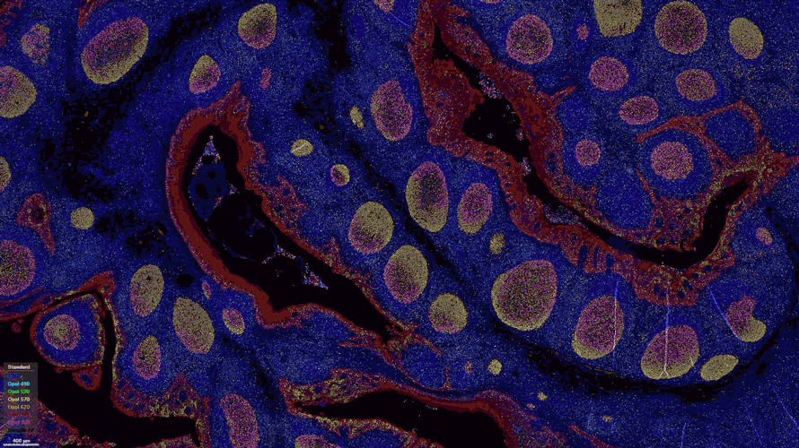
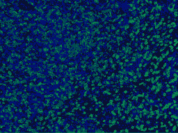
In immunofluorescence (IHC-IF) staining, antibodies that are bound to their respective proteins are conjugated with a fluorophore or fluorescent dye. To observe these proteins, a fluorescent microscope is used. Multiplex immunofluorescence, a subset of IF, utilizes various markers and wavelengths to detect multiple proteins with a single tissue sample.
Antibody validation is the process of confirming that an antibody specifically recognizes and binds to its intended antigen. Antibody optimization is used to enhance the performance of an antibody's characteristics through its sensitivity, and specificity. Both methods are used to ensure that antibodies are suitable for diagnostic and experimental applications.